Arteries In Neck Labeled - Subclavian Artery - You use your trapezius muscle for a variety of movements, including.. A ct angiogram of an elderly patient, obtained for evaluation of carotid stenosis, has been labeled in regard to both vessels and adjacent structures. Arteries of the neck model 9 photos of the arteries of the neck model arteries in the back of the neck, arteries in the neck and head, arteries veins neck, blocked arteries neck, clogged arteries neck, main arteries neck, major arteries neck, neck, arteries in the back of the neck, arteries in the neck and … anatomy face and neck side view The internal carotid artery, being one of the most clinically relevant and vital arteries, supplies oxygenated blood to crucial structures such as the brain and eyes. Tunica intima (the inner layer), tunica media (the middle layer), and tunica adventitia (the outer layer(2). These arteries arise in the neck, and ascend to the cranium.
Arteries and veins are a vital part of the circulatory system of all vertebrates. The vertebral arteries ascend through the neck inside the transverse foramina of the cervical vertebrae, all the way to the brain. In fact, the test may do more harm than good. Left and right common carotid. They supply blood flow to the brain.
Activity 2 Locating Arteries On An Anatomical Chart Or Model And Major Systemic Arteries Of The Body Flashcards Easy Notecards from www.easynotecards.com If one of them is narrowed or blocked, it can lead to a stroke. Tunica intima (the inner layer), tunica media (the middle layer), and tunica adventitia (the outer layer(2). In the neck, each carotid artery branches into two divisions: Instant anatomy is a specialised web site for you to learn all about human anatomy of the body with diagrams, podcasts and revision questions They supply blood flow to the brain. Labels include cephalic vein, brachial artery/vein, basilic vein, musculoskeletal nerve, ulnar collateral artery note the names of the major veins and arteries involved. The left common carotid comes directly off the aortic arch, while the right common carotid comes from the brachiocephalic. The cervical plexus is the main structure innervating or passing through the neck.
In the neck, each carotid artery branches into two divisions:
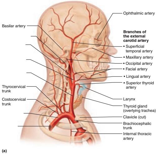
This section of the website will explain large and minute details of arterial anatomy of neck. As seen in the picture, it travels superiorly in a tortuous course along the under (ventral) surface of the tongue , below the longitudinalis inferior , and above the mucous membrane. In the neck, each carotid artery branches into two divisions: Left and right common carotid. They are the carotid arteries, and they carry blood to the brain. The external jugular vein (v. Through their course, they give off several meningeal, muscular and spinal branches for the nearby structures. There are two large arteries in the neck, one on each side. The superior gluteal artery and the inferior gluteal artery supply the gluteal region, but we'll take a look at the branches of the internal iliac artery in more detail in separate tutorials. The right common carotid artery has a different initial course. Arteries of the neck model 9 photos of the arteries of the neck model arteries in the back of the neck, arteries in the neck and head, arteries veins neck, blocked arteries neck, clogged arteries neck, main arteries neck, major arteries neck, neck, arteries in the back of the neck, arteries in the neck and … anatomy face and neck side view The main artery in the neck is the common carotid artery, which divides at the upper border of the thyroid cartilage of the larynx (c4). Each artery is a muscular tube lined by smooth tissue and has three layers:
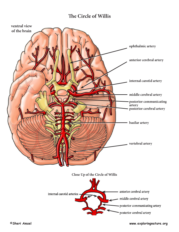
The left common carotid artery branches directly off the aortic arch and extends into the neck. Fig 5.14 blood flow to the head and neck processes of the cervical vertebrae where they unite to form a single basilar artery. Within the cranial vault, the terminal branches of these arteries form an anastomotic circle, called the circle of willis. The deep lingual artery (or ranine artery) is the terminal portion of the lingual artery after the sublingual artery is given off. In the neck, each carotid artery branches into two divisions:
Arteries Of The Head And Neck Advanced from www.exploringnature.org In fact, the test may do more harm than good. If one of them is narrowed or blocked, it can lead to a stroke. Arteries in the neck diagram, common carotid artery branches, external carotid artery function, how many carotid arteries, left common carotid artery function, the left common carotid artery supplies blood to the, what does the external carotid artery supply, what regions of the body are supplied blood by the external carotid artery, neck, arteries in the neck diagram, common carotid artery. Instant anatomy is a specialised web site for you to learn all about human anatomy of the body with diagrams, podcasts and revision questions The vertebral arteries are paired vessels which arise from the subclavian arteries, just medial to the anterior scalenes. The transverse cervical artery (also called the transverse artery of the neck) is a small blood vessel located in your neck. Labels include cephalic vein, brachial artery/vein, basilic vein, musculoskeletal nerve, ulnar collateral artery note the names of the major veins and arteries involved. Doctors can test for a narrowed carotid artery, but it's usually not a good idea.
The anatomy of neck arteries, normal variations, and anastomoses between different arteries is discussed in this chapter.
This artery provides blood supply to your trapezius muscle, a large muscle in your back that helps raise your arms. The vertebral arteries are paired vessels which arise from the subclavian arteries, just medial to the anterior scalenes. Arteries and veins are a vital part of the circulatory system of all vertebrates. There are two large arteries in the neck, one on each side. The cervical plexus is the main structure innervating or passing through the neck. Each artery is a muscular tube lined by smooth tissue and has three layers: (e.g., carotid arteries and jugular veins for the head). Fig 5.14 blood flow to the head and neck processes of the cervical vertebrae where they unite to form a single basilar artery. Carotid and vertebral arteries lie on either side of the neck(1). If one of them is narrowed or blocked, it can lead to a stroke. This becomes particularly important during investigation of head/neck vascular lesions. This artery provides blood to the right upper chest, right arm, neck, and head, through a branch called right vertebral artery. The right common carotid artery has a different initial course.
The basilar artery then terminates by dividing into two posterior cerebral arteries that supply the occipital and temporal lobes of the cerebrum. Common carotid artery the right and left common carotid arteries (cca) course superiorly on both sides of the neck lying within the respective carotid space, anteromedial to the internal jugular veins and accompanied by the vagus nerve. They ascend the posterior aspect of the neck, passing through holes in the transverse processes of the cervical vertebrae (known as foramen transversarium). This section of the website will explain large and minute details of arterial anatomy of neck. The cca courses superiorly in the neck, anteromedial.
Occipital Artery Neuroangio Org from www.neuroangio.org The basilar artery then terminates by dividing into two posterior cerebral arteries that supply the occipital and temporal lobes of the cerebrum. The right common carotid artery has a different initial course. The left common carotid comes directly off the aortic arch, while the right common carotid comes from the brachiocephalic. They supply blood flow to the brain. Fig 5.14 blood flow to the head and neck processes of the cervical vertebrae where they unite to form a single basilar artery. The transverse cervical artery (also called the transverse artery of the neck) is a small blood vessel located in your neck. In fact, the test may do more harm than good. Ninja nerds!join us in this video where we discuss the blood circulation of the head and neck using a flow chart.
The brachiocephalic trunk, left common carotid (cca), and left subclavian arteries.
The vertebral arteries ascend through the neck inside the transverse foramina of the cervical vertebrae, all the way to the brain. There are several head and neck arteries: They ascend the posterior aspect of the neck, passing through holes in the transverse processes of the cervical vertebrae (known as foramen transversarium). The vertebral arteries are paired vessels which arise from the subclavian arteries, just medial to the anterior scalenes. Doctors can test for a narrowed carotid artery, but it's usually not a good idea. This artery provides blood supply to your trapezius muscle, a large muscle in your back that helps raise your arms. This artery provides blood to the right upper chest, right arm, neck, and head, through a branch called right vertebral artery. The internal carotid arteries are branches of the common carotid arteries that bifurcate into the internal and external carotids at the level of the carotid sinus. A ct angiogram of an elderly patient, obtained for evaluation of carotid stenosis, has been labeled in regard to both vessels and adjacent structures. There are two paired arteries which are responsible for the blood supply to the brain; In fact, the test may do more harm than good. Carotid and vertebral arteries lie on either side of the neck(1). Arteries and veins are a vital part of the circulatory system of all vertebrates.
There are two carotid arteries, one on the right and one on the left arteries in neck. The vertebral arteries are paired vessels which arise from the subclavian arteries, just medial to the anterior scalenes.
Arteries In Neck Labeled - Subclavian Artery - You use your trapezius muscle for a variety of movements, including.. There are any Arteries In Neck Labeled - Subclavian Artery - You use your trapezius muscle for a variety of movements, including. in here.